
Teeth are hard, bone-like, and white, located in the lower and upper jaw bones. They are essential for functions such as chewing, biting, and speaking.
Teeth are of great importance in addition to being a bony-looking part of our body. It shows its vital importance in helping us chew, bite, laugh, and even talk in cooperation with the parts inside.
With its 3 main parts and 4 other parts, teeth are one of the most important parts of the human body.
Here’s a recap of the article about the important parts of a body:
- The crown, neck, and root are the main parts of teeth.
- Enamel, dentin, pulp, and cementum are the other parts of teeth.
- Teeth are not bone.
- Human teeth are 32.
- You can change the shape of your teeth as you want.
- Teeth affect speech.
- Your teeth can be transparent due to enamel loss.
- A healthy oral can be maintained by proper oral care with eating habits.
- Everyone has different teeth shapes.
- Your teeth can feel weird and many reasons can be behind it.
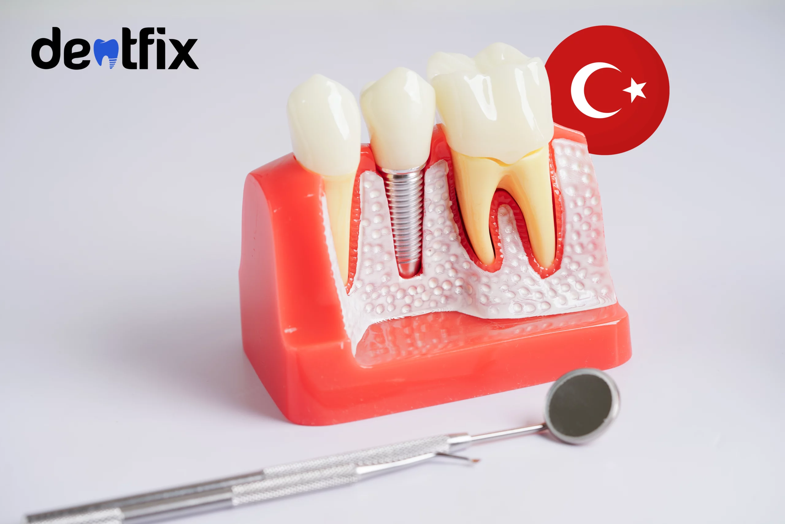
Tooth Anatomy
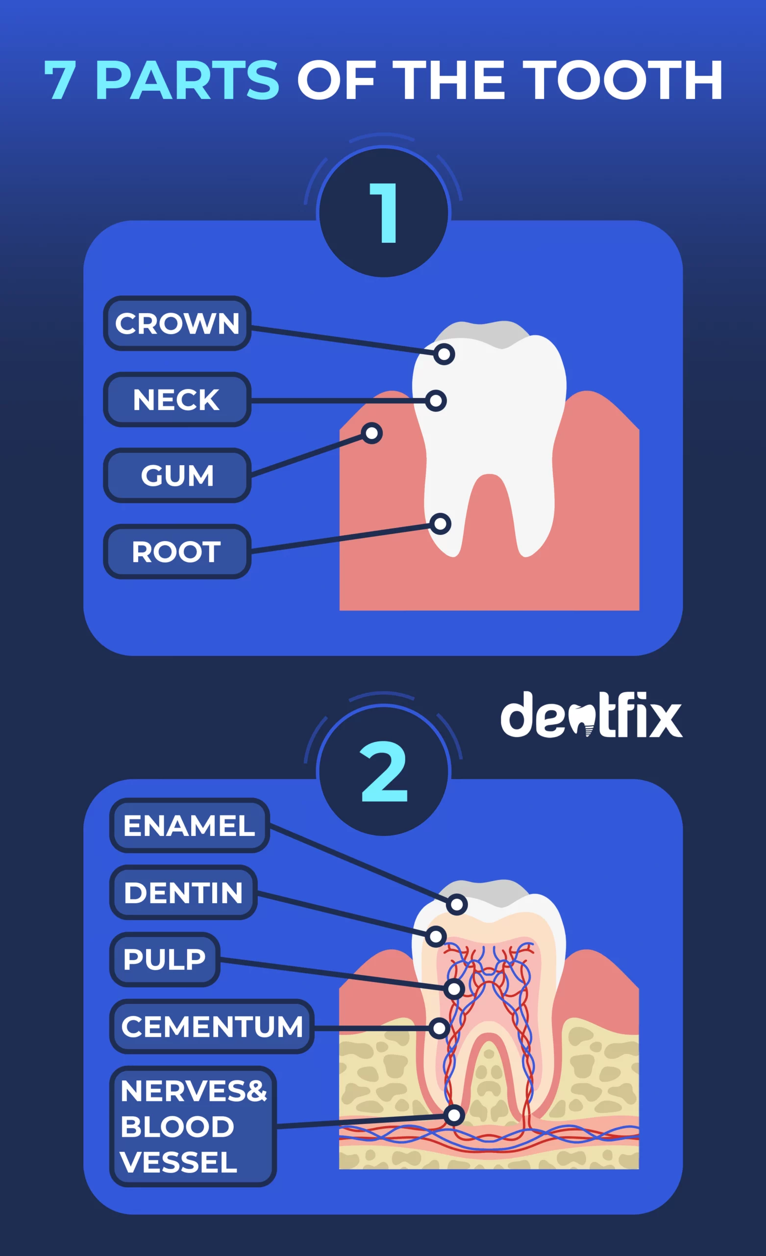
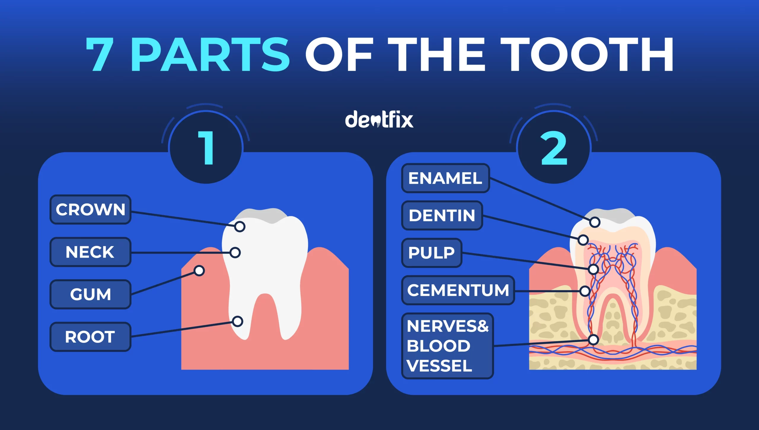
Tooth anatomy provides a detailed understanding of tooth structure. It consists of 3 main and 4 other parts of the tooth. They all have a function and work together.
The three main parts are:
- Crown: The crown is above the gum line and faces the oral cavity. It varies in function, such as incisors, canines, molars, and premolars. It plays a major role in speaking, chewing, and biting.
- Root: The tooth root is below the gum line and helps the tooth stay in place and withstand pressure.
- Neck: It is where the crown and root meet and form the line between the two layers of the tooth.
The four other parts are:
- Enamel: It covers the crown of the tooth. Enamel is the hardest layer of the tooth and the human body, protecting the tooth to a great extent.
- Dentin: It is under the enamel, is softer than the enamel, and has regrowth and regeneration properties.
- Pulp: It is under the dentin and consists of the dental pulp and root canals, and this is the innermost layer. It contains nerves, cells, and blood vessels.
- Cementum: It covers the root of the tooth. It has the feature of covering the root to the gums and jaw bones.
Apart from these, there are gums nerves, and blood vessels. Gum is rich in nerves and blood vessels protect the tooth root.
Is Tooth a Bone?
No. A tooth is not a bone.
Bone and teeth are considered the same because more than 99% of calcium can be found in the body and teeth. Another point is that they look similar and are both the hardest parts of the body.
They are not the same. There is only one difference between them. Bones can heal by themselves if they break. Teeth cannot heal, so they need to be protected.
Are Human Teeth 32 or 36?
Human teeth are 32 whereas babies have 20.
When they reach the age of six, their milk teeth will fall out and new teeth will grow. Then, they have 28.
When the molar comes out, the number reaches 32.
Adults can have 32 teeth if wisdoms are not extracted.
How Do Teeth Grow?
Teeth grow in generally four steps and these are:
Embryonic development: It appears as small buds on the gums, starting in the sixth week of pregnancy.
Formation of tooth structures: Tooth buds develop into the enamel, dentin, pulp, cementum, and periodontium structures.
Shedding of baby teeth: Baby teeth are shed to make room for permanent ones, starting around the age of 6.
Eruption of permanent teeth: Permanent teeth begin to emerge and this continues until wisdom teeth emerge.
Tooth formation begins before birth and continues until the emergence of permanent teeth (6-12 age). Tooth structures such as enamel, dentin, cementum, and periodontium must be formed to have a healthy tooth anatomy.
What Do Four Types of Teeth Look Like?
Each tooth is special, from person to person and from one another. There are four types of teeth. These four groups are incisors, canines, premolars, and molars.
They can be differentiated by being longer, shorter, pointed, or wider. Each of the teeth has a separate function.
| Positioning | Number | Function | Shape | Age | IMAGE |
Incisor teeth | front mouth | 8 | cutting | chisel-like | baby – 6 months adult – age of 6-8 |
|
Canine teeth | between incisors and pre-molars | 4 | tearing | sharp and pointy | baby – 16-20 months adult – 9-12 ages | 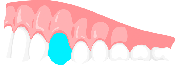 |
Pre-Molar teeth | behind canines | 8 | grinding, chewing | rectangular, 2-3 cusps | no baby premolars, adult – 9-13 ages | 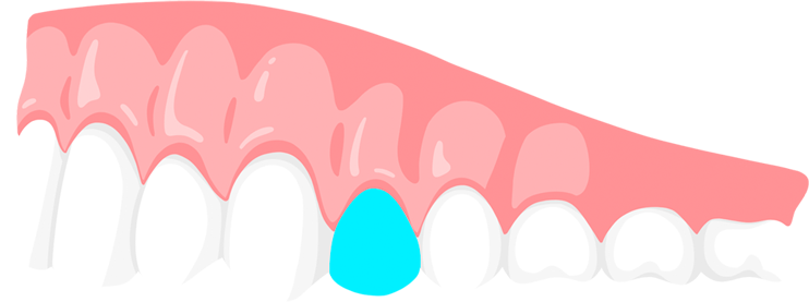 |
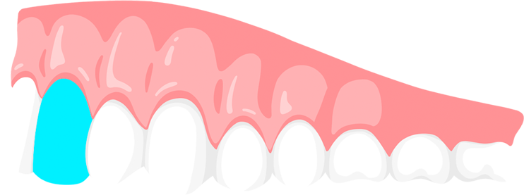
Molar teeth | back mouth | 12 | grinding, chewing | rectangular, 4-5 cusps, flatter surface | baby – 12-16 mouths adult – age of 6 | 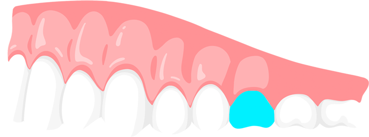 |
Types of Teeth Shapes in Dentistry
| Form | Distinctive features | IMAGE | |
| Aggressive style | Square | Flat teeth aligned in the same line |
|
| Dominant style | Square | Sharper and higher canines | 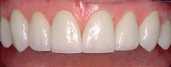 |
| Enhanced style | Slightly rounded edges | The height difference between central and lateral incisors, sharp canines | 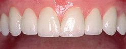 |
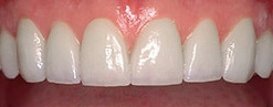
| Focused style | Similar to enhanced style except for central incisors | Central incisors are square, with no rounded edges | 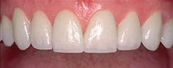 |
| Functional style | Similar to enhanced | Canines are pointier | 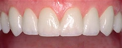 |
| Hollywood style | Square, similar to aggressive | Lateral incisors are slightly shorter than central incisors | 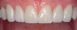 |
| Mature style | Square, similar to the aggressive style | Canines are more curved and slightly more pointed | 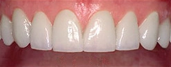 |
| Natural style | Similar to enhanced style | Canines are very sharp | 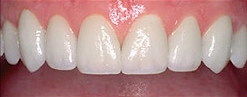 |
| Oval style | Oval tooth shape, curvy edges, and wider at center | Alignment is similar to the aggressive style | 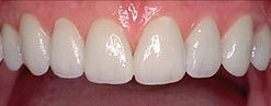 |
| Softened style | Similar to oval | Edges are less curved | 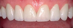 |
| Vigorous style | Square, similar to aggressive | Canines are longer, they protrude into the oral cavity | 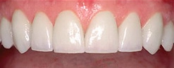 |
| Youthful style | Similar to the oval style | Canines are more protruding and sharp | 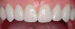 |
Can I Change the Shape of My Teeth?
Yes! You can change the shape of your teeth. There is a reshaping method called tooth filing.
The procedure is generally done for a cosmetic reason. It can be preferred for reasons such as preparing the tooth for operations and making room in the mouth.
As much enamel as necessary is removed and tooth shape is changed. After this process is completed, the tooth is polished and corrected.
| ⚠️ This procedure became increasingly popular and began to be performed at home. This is definitely harmful to teeth. If you are considering having this procedure done, you should consult a professional. |
The Stages of Dental Development
People have two types of teeth throughout their lives. First, we have baby teeth, then permanent teeth. The process in which these teeth are formed goes through certain stages.
Initiation Stage
It begins to form in the sixth or seventh week of the embryonic period. During this period, there is a developing tooth bud. The dental lamina connects this tooth bud to the epithelial layer of the mouth for a while.
Bud Stage
This occurs in the 8th week of the embryonic period. Cells called dental epithelium begin to bud. The cells will form the tooth embryo. This tooth embryo consists of the soft tissues necessary for the growth of a tooth.
Cap Stage
This is the stage when the outer layer of the tooth is shaped. The cells form a cap called an enamel organ that fits over the rest of the tooth bud. The reason why this head is called the enamel organ is that it will form the cells that produce enamel.
The tooth bud (Dental Papilla) forms the two inner layers of the tooth, these are dentin and pulp.
In addition, another cell called the dental follicle, which contains blood vessels and nerves, surrounds the enamel organ and the dental papilla, and thus the three elements of the tooth germ are on the scene. These are enamel, papilla, and dental follicles.
Bell Stage
The enamel organ takes the shape of a bell. Cells change, and so do their functions. Cells are the inner enamel epithelium, outer enamel epithelium, layer intermediate, and stellate reticulum, and they develop the enamel layer of the tooth.
Crown and Root Formation
Both the enamel and dentin are formed at this stage. These are the outer layers of the tooth.
The tooth root consists of a combination of three structures: the dental papilla, the dental follicle, and the root sheath.
Eruption Stage
The tooth has formed, and it is time for the final stage, which is called eruption. The formed tooth moves vertically above the gum line.
Baby teeth begin to appear at the age of six months and fall out around the age of 6.
What Is the Purpose of the Teeth?
The importance of teeth in life is undeniable.
Teeth make it possible to chew and digest food, however, it’s not just limited to that.
Teeth also play a big role in speech. It is key to forming sounds and speaking clearly when using words. While speaking, the tongue interacts with teeth, and as make sounds, teeth shape these sounds.
It shapes the face and supports facial length and the jawbone. Healthy teeth form a healthy and beautiful smile.
Which Teeth Play an Important Role in Speech?
Incisors in the mouth play an important role in speech. The two teeth are upper and lower, and the teeth beside them are incisors. They are the most visible teeth in the mouth.
When you use your words to speak, especially the front four incisors, they play a crucial role in making sounds. If they are missing, you will have difficulty pronouncing some sounds, such as ‘s’, ‘z’, ‘f’, ‘j’, ‘ch’, and ‘th’.
The teeth beside them don’t affect your speech. Only four, two upper and two lower, have a big effect on speech.
Your teeth may not touch, which means there can be a gap between the upper and lower teeth when you close your mouth. The reasons can be developmental and genetic factors or injuries. This is called an open bite and can lead to some issues, such as lisps or other types of speech issues.
| ⚠️ The American Dental Association (ADA) points out that this can be fixed before permanent teeth come. |
Does Missing Teeth Affect Articulation?
All teeth are important but some teeth can affect articulation.
Let’s see which one affects.
| Teeth Type | Place Of Teeth | Articulation |
| Missing Front Teeth | Upper and Lower | They affect your speech especially the sounds such as ‘s’, ‘z’, ‘f’, ‘j’, ‘ch’, ‘th’. |
| Missing Molar Teeth | Back of the Jaw | They don’t affect your speech. |
| Missing Premolar Teeth | Between Molars and Front Teeth | They don’t affect your speech. |
How to Maintain Dental Health
Maintaining oral care is key to maintaining dental health. Providing oral care involves making some routines permanent. It is necessary to make these a habit and do them without interruption.
These routines include brushing teeth, flossing, rinsing the mouth, and scraping the tongue.
It is necessary to do these at least two and at most three times a day. Brushing your teeth should last two minutes and rinsing your mouth should last 30 minutes. Thus, it strengthens the teeth against bacterial attack and prevents plaque formation.
Dental Care in Different Life Stages
Dental care is crucial in every life stage. You should maintain proper oral care to have healthy teeth and it should start at a very early age. The dental care routine can change a bit while aging.
| Life Stages | Routines | Differences |
| Children |
|
|
| Adults |
|
|
| Seniors |
|
|
The Connection Between Diet and Dental Health
There is a great relationship between the foods you choose and your teeth.
The foods you choose may not benefit your teeth and may, on the contrary, become the source of problems.
If you prefer acidic and sugary foods in your diet, it may mean that you are opening the door to tooth and gum problems. Acidic and sugary foods stick to plaque on the teeth and cause the teeth to be attacked by bacteria.
However, if you have a diet rich in calcium and protein, your teeth become stronger and healthier.
| Best Foods For Dental Health | Worst Foods For Dental Health |
| Cheese | Alcohol |
| Milk | Soda |
| Yogurt | Coffee |
| Carrots | Candies |
| Leafy Greens | Tomatoes |
| Apples | Pickles |
| Garlic and Onions | Juices |
| Nuts | Carbonated Drinks |
| Fish | Sport Drinks |
What Are the Common Dental Problems?
There are some common oral diseases. Here is their prevalence:
- Tooth decay: 3.5 people suffer from tooth decay globally according to the World Health Organization. (WHO).
- Gum disease: Centers for Disease Control and Prevention (CDC) point out that over 47% of people have gum diseases.
- Tooth sensitivity: The International Dental Journal stated that one out of eight people have tooth sensitivity.
- Oral cancer: According to the World Cancer Research Fund, it occurs in 150,000 people every year.
- Bad breath: It is found in approximately 25% of people according to the International Journal of Oral Science.
- Malocclusion: According to The World Health Organization (WHO), prevalence varies from 39% to 93%
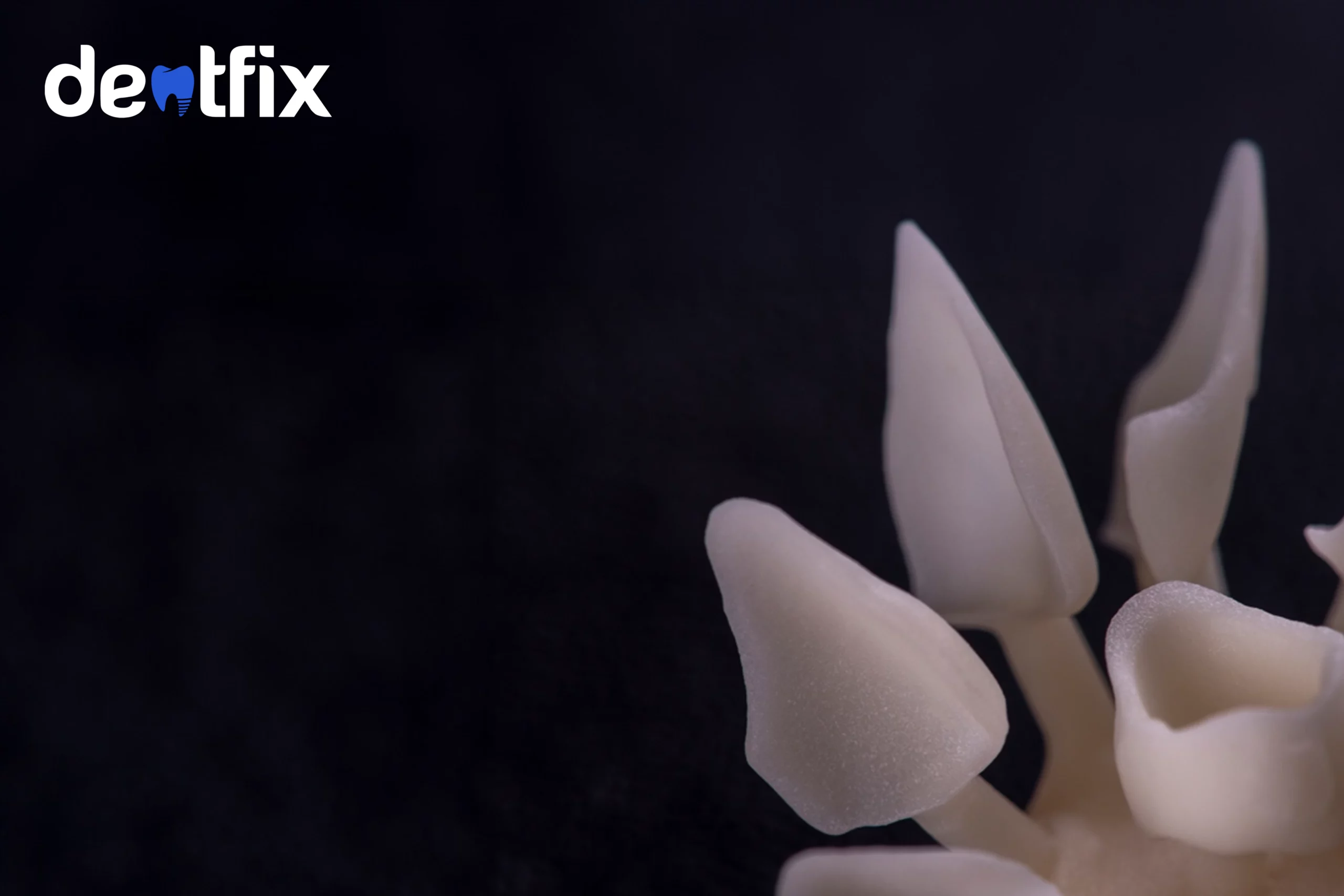
Why Is My Teeth Transparent?
Transparent teeth are an indication of enamel loss. For various reasons, the outermost hard enamel layer of the tooth can become thin and eroded. Over time, teeth can be in great danger due to tooth enamel loss. So, if you’re wondering why is my teeth transparent, the answer is all about the enamel.
The reasons can be:
- Consuming acidic and sugary nutrients
- Celiac disease
- Harshly brushing teeth
- Dry mouth
- Gastrointestinal problems
- Medications
How to Fix Transparent Teeth
There is a solution to save transparent teeth. Solutions aim at protecting the tooth against damage that may occur as a result of enamel loss.
- Fluoride treatments: This is used to strengthen teeth and enamel from further erosion.
- Dental bonding: A tooth-colored resin is applied to the transparent tooth which is caused by enamel loss and hardened with a special light.
- Dental crown: Enamel loss can be severe, in which case a cap covering the tooth, a crown, is placed on the tooth.
- Root canal: If enamel loss is severe, the nerve of the tooth may be exposed, in which case root canal treatment is performed to protect the tooth from further damage.
Does Your Teeth Feel Weird?
Sometimes your teeth may feel weird. When you check your teeth, you may not notice anything, but you may wonder why is my teeth feel weird. Teeth are sensitive and the nervous system can send signals to the teeth if the reason is:
- Malocclusion: This is a type of biting that means that your upper and lower teeth do not fit and close properly. This makes your teeth feel weird while biting or chewing.
- Gum disease: When gums become inflamed, your tooth may feel strange. Redness and swelling occur in the gum, causing an uncomfortable feeling in the tooth.
- Oral care ingredients: If you are allergic to the ingredients in the materials you use in your oral care routines and if they cause dry mouth, this can lead to a weird feeling.
- Food stuck between teeth: Sometimes food can get between the teeth and its presence there can cause an uncomfortable and weird feeling in teeth and even gums.
Why Do My Teeth Feel Weird After Eating Spinach?
Saliva contains calcium crystals. Spinach contains oxalic acid. When spinach is eaten, the calcium crystals in the saliva meet with the oxalic acid in the spinach. Thus, calcium oxalate crystals are formed.
This combination is insoluble in water and sticks to the tooth. Your teeth may feel weird because it is an unusual combination and sensation.
How to Deal with Dental Emergencies
Conditions such as severe tooth decay, inflammation, chips and cracks in the teeth, tooth abscess, teeth fracture, and loss of crowns may require a dental emergency.
When faced with this situation, you should consider three options.
Your first thought should be to call your clinic. If they are not available, reaching your nearest dental emergency clinic is the second option. Since there are neither, you can choose regular emergency rooms.
Cosmetic Treatment Options for Dental Problems
There are various cosmetic treatments for dental problems.
If you have chipped or cracked teeth, if you have gaps between your teeth, if you want to change the shape of your teeth, or if you are not satisfied with the color of your teeth, there are treatment options for all of them. These treatment options aim to make your teeth look healthier and better:
- Veneers
- Dental Crowns
- Dental Implants
- Dentures
- Hollywood Smile
- Teeth Whitening
- Dental Contouring
Fun Facts about Teeth
There can be some fun facts about teeth.
- Teeth are so unique that dental records are used for identification.
- Tooth enamel made up of 89% of calcium is harder than bone.
- A healthy tooth can withstand 5600 pounds of pressure per square inch.
- The first dentist was an Egyptian person who lived in 2600 BC.
- Teeth have a memory and tend to return to their original position when braces are removed. That’s why you need to use a retainer after braces.
- Since the density levels of the inner and outer parts of the teeth are different, sudden temperature differences create stress.
- People used animal teeth as false teeth in old times.
- Genetic factors, ethnicity, and dietary habits can influence teeth.
- Pointed and elongated teeth were associated with social status in some societies.
References:
1) Arola DD, Gao S, Zhang H, Masri R. The Tooth: Its Structure and Properties. Dent Clin North Am. 2017 Oct;61(4):651-668. doi: 10.1016/j.cden.2017.05.001. PMID: 28886762; PMCID: PMC5774624.
2) Pashley DH. Dentin: a dynamic substrate–a review. Scanning Microsc. 1989 Mar;3(1):161-74; discussion 174-6. PMID: 2662395.
3) Kinney JH, Marshall SJ, Marshall GW. The mechanical properties of human dentin: a critical review and re-evaluation of the dental literature. Crit Rev Oral Biol Med. 2003;14(1):13-29. doi: 10.1177/154411130301400103. PMID: 12764017.